
Laura Vittadello | University of Osnabrück | Osnabrück, Germany
“Tunable High Energy Microscope: Concept, Realization and Growing Field of Applications in life sciences”
Scientific Talks, Session II | Monday, September 12 | 14:10 – 14:40

Nonlinear optical (NLO) microscopy has emerged as successful tool in the last years within the bio-medical research for the possibility to perform imaging of intact live organisms. Its success resides in the ability to image deeper, obtain 3D data, decrease the out of focus background and restrict the photodamage to the focal point. On the other side the exploitation of NLO effects requires the use of high optical peak intensities, usually achieved by focusing the laser through a microscope objective or via complex laser shaping. Our approach to this problem consists in a re-thinking of the laser space parameters in a region of high energy per pulse, low repetition rate in the kHz regime and wavelength up to 2200nm, in counter-trend with the actual microscope research technology. This is technologically realized via the exploitation of regeneratively amplified fs-laser and optical parametric amplifiers, representing the core of the new proposed Tunable hIGh EneRgy (TIGER) nonlinear microscope [2].
This redesign allows easily to conceive a widefield nonlinear microscope (Fig 1a). Indeed, it is now possible to employ nonlinear effects in an area up to 1.5×1.5mm without complex laser shaping, recording dynamical processes with frame rates up to 100 fps. The impact of the new parameters as the energy per pulse, the pulse repetition rate as well as of the photon energy is evaluated via, harmonic nanoparticles, here chosen as biological markers because of their high nonlinear optical coefficients [3], absence of blinking and good biocompatibility. All these concepts are then applied to the nanoparticles tracking via second harmonic generation in the heart of a living Drosophila (Fig. 1b), providing experimental access to the velocities of the hemolymph flux.
The new setup opened also the possibility to use easily a wavelength regime poorly exploited but in principle beneficial for deep imaging, i.e up to 2200nm [4] and to induce cell membrane lesions with nanometric precision [5].
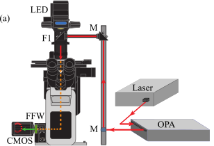
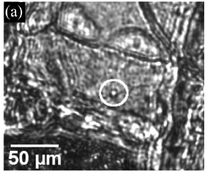
Figure 1: : (a) Sketch of front view of the TIGER microscope. A regenerative amplified fs-laser acts as a source for an optical parametric amplifier (OPA) which in turn is coupled into an inverted microscope frame via a periscope. The light emitted by the sample is collected by an objective, filtered from the fundamental pulse radiation and redirected onto a CMOS camera. (b) Zoom on the heart structure where an HNP flowing in the heart chamber is visible in the white circle. Taken from Ref. [5].
References
[2] Vittadello, L. et al. Opt. Mater. Express 2021, 11, 1953.
[3] Bonacina, L. Molecular Pharmaceutics, 10(3):783–792, nov 2012.
[4] Vittadello, L. et al. Nanomaterials 2021.
[5] Niekamp, P. et al, Nat Commun 13, 1875 (2022).