
Tomáš Čižmár | Leibniz IPHT | Jena, Germany
“Hair-thin holographic endoscope for neuroscience advances”
Scientific Talks, Session IX | Wednesday, September 14 | 12:10 – 12:40

Imaging in extended depths of brain tissue, whether non-invasively or invasively, is associated with numerous limitations. Most importantly, non-invasive approaches suffer from steep quality loss with increasing depth, while invasive alternatives (endo-microscopes) cause profound tissue damage, particularly in the vicinity of the structures under investigation.
About a decade ago, a new technological candidate emerged, with the potential to deliver sub-micrometre-resolution images through optical fibre, as narrow as a human hair (~0.1 mm), with no additional optics at its distal end required. Unlike common optical systems, built around predictable imaging components like lenses, the hair-thin endoscope involves an optically complex medium in the form of highly multimode optical fibre, which transports light between its extremities in a seemly-random and a priori unknown manner. The young and rapidly growing discipline of complex media photonics devised methods based on digital holography as a solution to how such complex light transport can be experimentally quantified and utilised for imaging, thereby turning a single fibre into an ultra-thin analogue of a microscope objective. Importantly, these new so-called holographic endoscopes provide diffraction-limited observations with the optimum performance, regardless of the fibre probe length, making them applicable in unprecedented tissue depths.
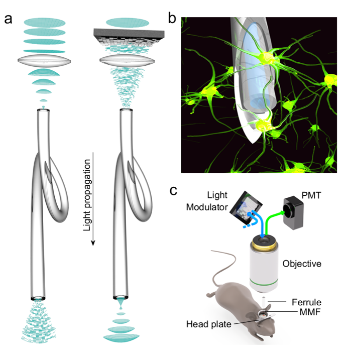
Fig. 1: Holographic endoscopy. (a) Coherent light propagated through a MMF forms a speckle pattern at the output (left). Manipulation of the wavefront entering the MMF yields a desired optical field, e.g. a focal spot (right). (b, c) Experimental configuration for in vivo imaging.
In the talk, I will discuss the most severe limitation of image degradation due to fibre bending [1], and various applications including optical trapping [2], and 3D imaging of macroscopic objects [3].
In the context of neuroscience [4], I’ll showcase our most powerful, highly optimised and stable instrument across a large spectrum of possible applications including structural sub-micrometre resolution imaging in deep brain regions (amygdala), intracellular calcium imaging and single-vessel blood flow velocity measurements.