


Charity Hayes Rounds
Assessment of a Fresnel biprism-based digital holographic microscope for fast, high-sensitivity, high-resolution and polarization-sensitive phase imaging
The University of Memphis // Memphis, USA
Q&A Session I // Wednesday, October 38 // 6.15 pm – 7 pm (CET)

Among all quantitative phase imaging (QPI) techniques, digital holographic microscopy (DHM) is one of the most promising to perform label-free live-cell imaging while providing topographical information on the specimen. Alternatively, DHM provides five key advantages of stability, speed, accuracy, spatial resolution and polarization sensitivity that have not been attained in other QPI methods. In this work, we present a common-path polarization-sensitive (PS) DHM system based on the use of a Fresnel biprism (FB) [1]. The proposed DHM system offers the five desired features in QPI simultaneously: high temporal sensitivity, high speed, high accuracy, high spatial resolution, and sensitivity to birefringence. To the best of our knowledge, this is the first FB-based DHM system providing these five features altogether.
The optical configuration of the proposed common-path DHM system operates using a briprism illustrated in Figure 1. And the major component of the proposed system is inserting the FB between the tube lens (TL) and the sensor plane. Thus, at any observation plane behind the FB, there is a coherent superposition between the two split beams generated by the biprism. Sinusoidal fringes are generated in the region of superposition and its maximum lateral extension is given by L/2 where L is the biprim’s lateral extension. It is important to mention that, in this common-path DHM system, one only reconstructs the phase information that it is encoded within the fringes’ FOV (e.g. cone-shape region).
The benefits of assembling all five features of high speed, high resolution, high accuracy, high stability and polarization sensitivity in the proposed system are exhibited by imaging a monolayer of human U87 glioblastoma cells. Figure 2(a) shows the phase information from U87 glioblastoma cells. To provide polarization-sensitive (PS) measurements, the sample is illuminated with linear polarized light whose plane-of-vibration is varied by rotating a linear polarizer inserted before the sample holder in Fig. 1 (not shown). Figure 2(b) shows the phase histograms of the phase image from U87 glioblastoma cells illuminated with two different polarization angles. From the distinctive behavior of the cells at different polarization angles, we have demonstrated their anisotropic properties.
We have implemented a FB-based common-path DHM system to achieve accurate quantitative PS phase images in a new, simple, compact, and cost-effective way, recognizing the low cost (a few hundred dollars) associated in implementing this simple architecture, enabling the use of this QPI technique accessible to most laboratories with standard light microscopes.
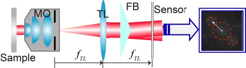
Fig. 1. Proposed common-path DHM using a Fresnel biprism (FB). The system operates in off-axis regime.
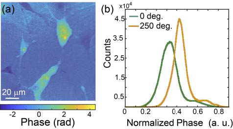
Fig. 2. (a) Pseudocolor phase image of human U87 glioblastoma cells. (b) Normalized histogram of the phase images of the cells at
Further Talks